facs buffer flow cytometry
Ad Search over 300000 products and 500 service solutions for your research needs. Flow cytometry FACS staining protocol Cell surface staining 1.
Flow Cytometry Perm Buffer 10x Pf00011 C Proteintech
Ad Includes One Bottle Of FCM Lysing Solution FCM Wash Buffer More.
. We use this buffer for surface staining as well as for intracellular staining. Request a quote and see how Agilent has advanced the boundaries of flow cytometry. Resuspend cells with 052 mL FACS buffer.
1 Centrifuge as above fixed cells and resuspend pellet in 1 ml of PBS. Gain an Unparalleled Understanding of the Spatial Relationships Between Cell Types. Incubate on ice for 30-60 minutes in the dark.
Flow Cytometry Staining Buffer FACS Buffer This basic FACS Buffer is a buffered saline solution that can be used for immunofluorescence staining protocols antibody and cell dilution. FACS Buffer we use has 1 BSA and 01 Sodium Azide. BD FACS Sample Prep Assistant SPA III.
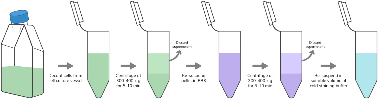
Harvest wash the cells single cell suspension and adjust cell number to a concentration of 1-5106 cellsml in ice cold FACS. Ad Imaging Mass Cytometry - The Most Proven Approach to High-Multiplex Imaging. Our FACS buffer is based on PBS and contains 2 FCS 005 Sodium Azide.
Also compare to BioLegend buffers to the equivalent BD products. The Click-iT EdU Flow Cytometry Assay Kits are novel alternatives to the BrdU assay. Wash 1-3 times as described throughout this protocol.
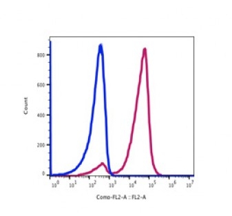
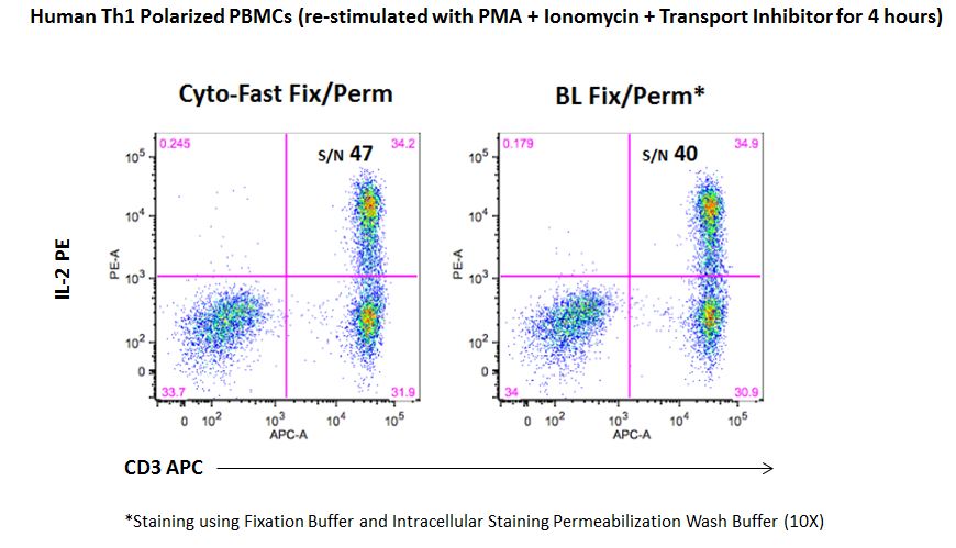
When compared with cells stained without a Brilliant Stain Buffer Left Plots. Place samples in 12 x 75 mm Falcon tubes and analyze by flow cytometry as soon as possible within 1 hour. Alternatively samples can be.
Resuspend cells in an appropriate volume of staining buffer with care to avoid. Ad Learn how Agilent NovoCyte Flow cytometers have advanced the boundaries of flow cytometry. Add either 100 µl for microwell plates or 250 µl for tubes aliquots of fixation buffer to each cell pellet and resuspend the cells by either pipetting or vortexing.
This Flow Cytometry Staining Buffer is a buffered saline solution containing fetal bovine serum and sodium azide 009 as a preservative. EdU 5-ethynyl-2-deoxyuridine is a nucleoside analog to thymidine and is incorporated into DNA. View available buffers for various flow cytometry applications.
BSA and FBS or any other serum for that matter will accomplish pretty much the same thing when staining cells for flow. This buffer can be used for antibody and cell. 2 Add 100 μl of 200 μgml DNase-free RNaseA and incubate at 37C for 30 minutes.
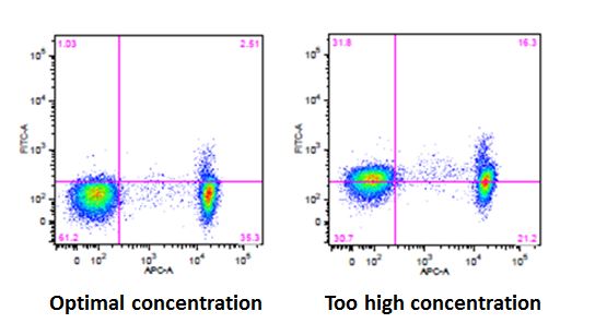
BioLegend develops and manufactures world-class. The fluorescence of the fluorochrome has faded. If titrating antibodies and storing aliquots of the same add sodium azide in the storage buffer at 009.
Gain an Unparalleled Understanding of the Spatial Relationships Between Cell Types. Staining buffer is the buffer used. Ad Confirm verify and optimize your gated cell populations with real-time image tracking.
Ad Imaging Mass Cytometry - The Most Proven Approach to High-Multiplex Imaging. Run sticky samples at high flow rates with a system that is less sensitive to clogging. Be sure to store the conjugated.
Flow cytometric analysis was performed using a BD. 3 Add 100 μl of 1 mgml. Ad Browse Discover Thousands of Science Book Titles for Less.
Offers a Range Of Blocking Reagents For Use In Western Blotting Research Applications.

Brilliant Stain Buffer

Functional Flow Cytometry Of Monocytes For Routine Diagnosis Of Innate Primary Immunodeficiencies Journal Of Allergy And Clinical Immunology

Fluorescence Activated Cell Sorting Facs Sino Biological
Stain Buffer Fbs

A Flow Cytometry Based Assay For Serological Detection Of Anti Spike Antibodies In Covid 19 Patients Star Protocols

Flow Cytometry Protocols
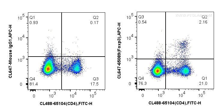
Facs Buffer Composition
Ebioscience Flow Cytometry Staining Buffer
Blog 3 Considerations For Intracellular Flow Cytometry Icfc
Blog 3 Considerations For Intracellular Flow Cytometry Icfc

Fluorescence Activated Cell Sorting Of Live Cells Abcam

Protocol For High Throughput Compound Screening Using Flow Cytometry In Thp 1 Cells Star Protocols
Facs Buffer Composition

What Is Flow Cytometry Technology Networks

Fixation Buffer
Flow Cytometry Sample Preparation

Flow Cytometric Characterization Of Tissue Resident Lymphocytes After Murine Liver And Heart Transplantation Star Protocols

Flow Cytometry Facs Protocols Sino Biological

Pre Sort Buffer